TMT
TMT test is a stress test that checks the functioning of the heart while the person is exercising on a treadmill. The test measures the blood circulation when the person is resting and when under optimum physical pressure. Abnormal heartbeats during strenuous physical activity indicate the presence or absence of coronary artery diseases. The procedure needs a person to move on the treadmill. The electrical activity of the heart gets recorded. The speed and slope of the treadmill are changed at intervals to see how heart circulation varies to different levels of exercise. There is regular monitoring of blood pressure, heart rate. Experts at regular intervals inquire from the person about the symptoms such as chest pain, dizziness and unusual shortness of breath, or extreme fatigue if he experiences. Test facilitates detecting congenital heart problems, determining the condition of a person after angioplasty or heart attack. Repressed conditions like Shallow breathing, dizziness, chest discomfort also get detected. The wide-scale applicability of TMT in determining various heart conditions helps to determine the probability and extent of coronary disease.
ECHO
ECHO test makes use of sound waves to generate images of the heart. It helps doctors check the functioning of valves and chambers, detect congenital heart defects before birth.
ECHO facilitates the detection of damage to the heart muscles and other heart defects. The procedure needs a person to lie on the bed and undress from waist up.
Technicians attach sticky patches known as electrodes to the body that help detect and conduct electrical currents. Applying gel to the transducer helps improve the conduction of sound waves. It helps in generating clear images of heart circulation. Technicians do recordings and frame reports that the concerned doctor evaluates further.
EEG
EEG is a test that uses small metal discs to detect electrical activity in the brain. EEG recording shows wavy lines that are electrical impulses sent by the brain all the time. The test plays a vital role in diagnosing brain disorders, and it also helps confirm brain death if a person is in a persistent coma. It also helps experts decide the right amount of anaesthesia for a medically induced coma. The technician marks the points on the head to fix electrodes. The gel or cream is applied to these marks to improve the quality of the recording. Special adhesives or electrode caps help attach electrodes to the scalp. Amplify instruments connected to Electrodes amplify brain waves and record them on computer equipment. The technician may ask the person to perform simple activities while he monitors the brain activity. A person may need to do a few simple calculations, read a paragraph, look at a picture, breathe deeply for a few minutes, or look at a flashing light. Technician recording the test prepares reports, and doctors interpret the recording and discuss the results with the patient.
ECG
ECG is a recording of electrical signals in the heart. It helps detect heart problems and monitor heart health. Experts recommend ECG if they hint blocked or narrow arteries are a cause of chest pain. Abnormal heart rate also arise the need for ECG. The test helps to diagnose the efficiency in working of the pacemaker. ECG test involves attaching electrodes to the chest and limbs. Electrodes stick to the body on one end, and on another side, they have wires that connect to a monitor. Electrical signals made by the heart get recorded on the computer. On the screen, the waves appear that technicians observe and prepare reports. Experts analyze the ECG reports and discuss the results with the patient. They talk about the abnormalities they encounter in heart rate, rhythm, structural abnormalities, or doctors examine inadequate blood and oxygen supply to the heart.
X-ray
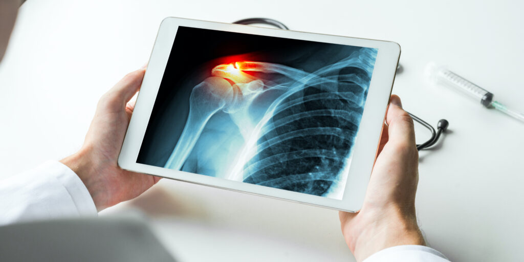
The most common imaging test used for decades is the X-ray. It helps experts visualize the area where a person reports pain or discomfort, monitor the progression of a diagnosed disease or check the efficiency of the treatment prescribed. There are no standard procedures for taking an X-ray. Technicians recommend the posture and positioning of the body based on the region of the body that needs an X-ray. X-ray uses a small amount of radiation to generate an image that is safe for adults. In some cases, there may be the need to take contrast medicine before an X-ray. The radiologist collects the X-ray images, prepares the report, and hand-over them to the concerned doctor for consideration and treatment.


